
“You can tell so much about the basic composition of a material by looking at its diffraction pattern” Prof Nigel Allinson MBE. Distinguished Professor of Image Engineering. University of Lincoln and one of the founders of ISDI.
X-ray Crystallography is a scientific technique which owes much of its development to the work of Lawrence Bragg.

Who was Lawrence Bragg?
Lawrence Bragg, along with his father, William Henry Bragg, played a pivotal role in establishing X-ray crystallography. In 1912, while a student at Cambridge University, Lawrence Bragg made a crucial breakthrough by interpreting the diffraction patterns produced when X-rays passed through crystals. He realized that these patterns revealed the atomic structure of the crystal.
In 1915, at just 25 years old, he was awarded the Nobel Prize in Physics alongside his father, making him the youngest physics Nobel laureate to date. Their work laid the foundation for a technique that would revolutionize our understanding of matter.
What is X-ray Crystallography?
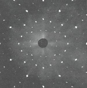
X-ray diffraction image
X-ray crystallography is the scientific method used to determine how atoms are arranged in a crystal. X-ray crystallography does this with X-ray radiation.
Crystalline Materials: Crystals are materials where atoms are arranged in a highly ordered, repeating pattern.
X-ray Interaction: When X-rays are directed at a crystal, they interact with the electrons of the atoms, causing them to scatter in a manner that is determined by the structure of the atoms.
Diffraction Pattern: The scattered X-rays create a unique diffraction pattern, a series of bright and dark spots, which is captured on film, imaging plate or with a digital X-ray detector.
Determining Structure: By analysing this diffraction pattern, scientists can determine the positions of the atoms within the crystal and thus understand the molecular structure.

The underlying physics of this process is explained by Bragg's Law, which describes how X-rays interact with the atomic planes in a crystal. It relates the angle at which X-rays strike the crystal to the distance between the planes of atoms. The law is expressed by the equation:
nλ = 2d sinθ
Where:
n is an integer representing the order of reflection
λ is the wavelength of the incident X-rays
d is the distance between the crystal’s atomic planes
θ is the angle of incidence (and reflection) of the X-rays
The Impact of X-ray Crystallography
X-ray crystallography has had a profound impact on various scientific fields:
Chemistry: It has allowed scientists to determine the structures of countless molecules, including pharmaceuticals and catalytic converters, leading to advancements in drug development and materials science.
Understanding the three-dimensional structures of DNA and proteins and has been crucial for deciphering biological processes and developing new therapies. Arguably, the most famous X-ray crystallography image was ‘Photograph 51’ acquired by Rosalind Franklin in May 1952 at King’s College London, which led directly to the discovery of the structure of DNA by Penrose and Crick in Cambridge in 1953.

‘Photograph 51’ - read our post ‘Celebrating Rosalind Franklin's Contribution to the Discovery of DNA's Structure’
Materials Science: It helps in designing new materials with specific properties, such as stronger alloys and more efficient semiconductors.
Mineralogy and Geology: It identifies and characterizes minerals and rocks, providing insights into the Earth's composition and history.
Using CMOS in X-ray Crystallography
In crystallography, CMOS X-ray detectors and image sensors, are used to record diffraction patterns from crystals. Protein crystallography has been an area of research that benefited from advances in X-ray detectors using first fibre coupled CCDs with gadox scintillators and more recently CMOS images sensors and direct conversion, photon counting X-ray detectors.
How they work:
X-rays, generated by a synchrotron or conventional home lab source, interact with the crystal, causing the X-rays to be diffracted.
The diffracted X-rays then interact with a scintillator and light to be emitted. The light pattern is recorded by a CMOS image sensor, creating a digital image of the diffraction pattern.
The digital image is then processed and analysed using specialist software.
How is X-ray crystallography used in the world today?
Today, X-ray crystallography continues to be an essential tool in scientific research. Synchrotron radiation sources, large and expensive facilities which produce extremely intense and coherent X-ray beams, have enabled the determination of highly complex structures. Home lab sources and modern digital X-ray detectors have made crystallography techniques widely available in academic and commercial labs as well as at synchrotron radiation facilities.
William Lawrence Bragg's work opened a window into the atomic world, allowing us to see the building blocks of life and materials. Although electron microscopy has become the dominant analytical modality in biology in recent years, X-ray crystallography remains a powerful and indispensable tool in modern science, continuing to reveal the secrets of the universe at the atomic level.
Coming soon, an interview with founders of ISDI Professor Nigel Allinson discussing the use of CMOS in X-ray powder diffraction, which can be used for quality control when testing drugs in developing countries.